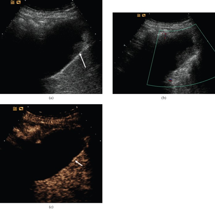
Figure 1.
Biliary sludge. (a) Baseline US image depicts echogenic material within the gallbladder (arrow). This does not exhibit movement on change in patient posture. (b) Colour Doppler US demonstrates no noticeable vascularity. (c) Late arterial-phase CEUS obtained 30 s after administration of microbubble contrast demonstrates that there is no enhancement of the echogenic material, i.e. the contents of the gallbladder remain dark, indicating that it is non-vascular and simply represents adherent biliary sludge. The gallbladder wall enhances (arrow) and is seen separately from the lesion, confirming no mural abnormality.

