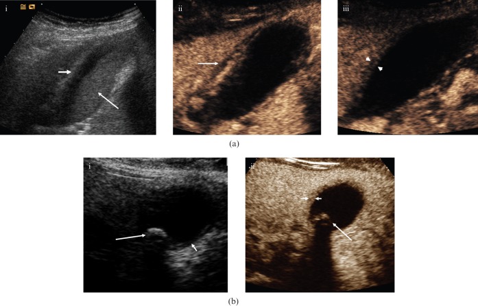
Figure 2.
Acute cholecystitis. (a) (i) Baseline longitudinal US of a 37-year-old female depicts a moderately distended gallbladder, hazy delineation of the thickened (>3 mm) gallbladder wall (small arrow) and echogenic debris within the gallbladder (large arrow). (ii) CEUS 30 s after administration of microbubble contrast depicts an initially hypervascular gallbladder wall. There is improved delineation of pericholecystic fluid (arrow). Note the lack of enhancement of the echogenic sludge within the gallbladder. (iii) CEUS at 86 s. In the late phase, the gallbladder wall shows less enhancement and is hypovascular (between arrowheads) relative to the adjacent liver parenchyma. This enables more accurate measurement of wall thickness. (b) (i) Axial baseline US image of a 30-year-old female with acute cholecystitis; thickening of the gallbladder wall (short arrow) and gallstones are present (long arrow). (ii) CEUS at 80 s shows the differential enhancement of the gallbladder wall (short arrows), allowing accurate assessment of mural thickness. Note the lack of enhancement of biliary sludge and the clear demarcation of the solitary calculus (long arrow).

