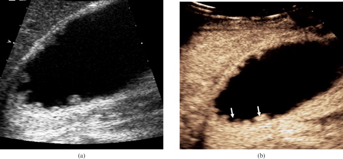
Figure 3.
Adenomyomatosis. (a) Longitudinal baseline US shows segmental mural thickening in a 37-year-old asymptomatic male patient in keeping with a diagnosis of adenomyomatosis. (b) CEUS 23 s after administration of microbubble contrast demonstrates enhancement of the thickened gallbladder wall with anechoic diverticula (arrows).

