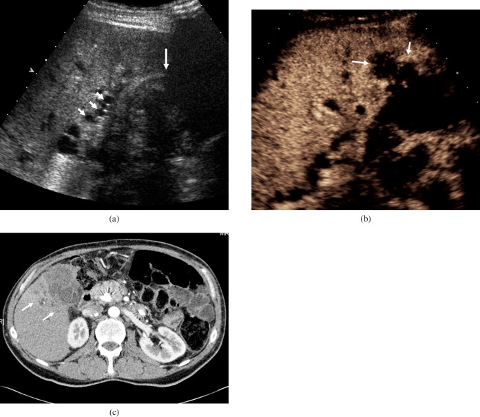
Figure 6.
Adenocarcinoma of the gallbladder. (a) Baseline US in a 57-year-old man presenting with obstructive jaundice depicts a soft-tissue mass in an ill-defined gallbladder (long arrow) and evidence of bile duct dilatation (short arrows). (b) CEUS image obtained 90 s after administration of microbubble contrast demonstrates a hypovascular mass centred on the gallbladder fossa, with invasion into the hepatic parenchyma (arrows). The late-phase hypovascularity suggests malignancy and differs from the persistent enhancement in cholecystitis. (c) Axial contrast-enhanced CT at the level of the gallbladder fossa demonstrates a mass with invasion into the adjacent liver parenchyma (arrows), appearances that correlate well with those seen at CEUS.

