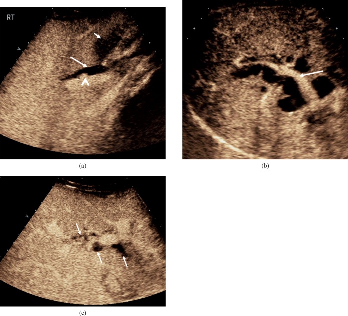
Figure 7.
Biliary duct dilatation. (a) The common hepatic duct is dilated (long arrow) in this 28-year-old female following recent passage of a biliary calculus. Enhancement is seen in the hepatic artery adjacent to the duct (arrowhead). In addition, inflammatory changes in the gallbladder wall are evident (short arrow). (b) Intrahepatic duct dilatation secondary to carcinoma of the head of the pancreas in a 78-year-old male. The portal vein demonstrates enhancement (arrow) with the surrounding dilated bile ducts demonstrating no enhancement. (c) Focal biliary dilatation (arrows) in the graft liver following liver transplantation in a 52-year-old male with an occluded hepatic artery. Ischaemic changes in the bile ducts are probably responsible for this appearance.

