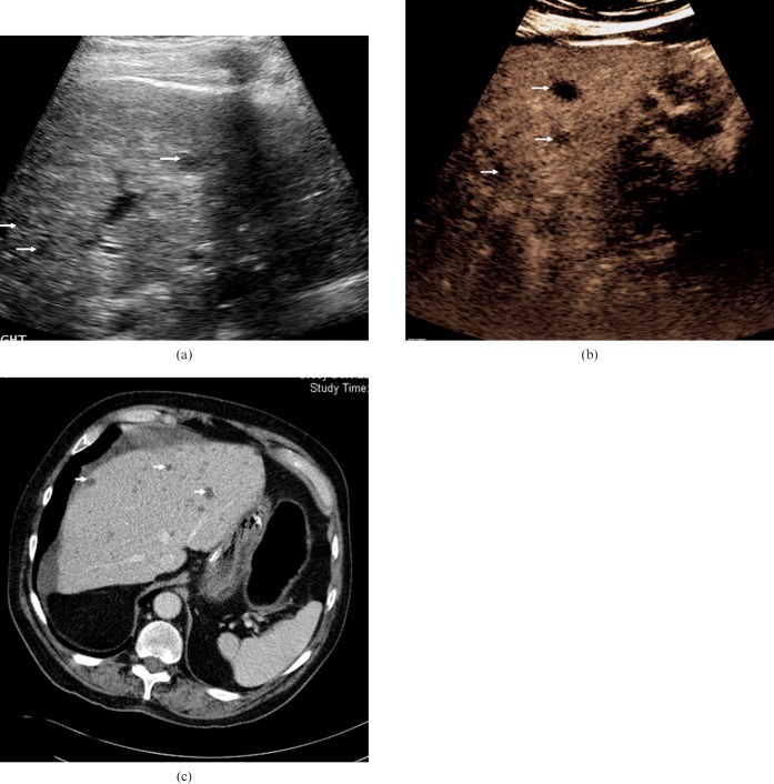
Figure 9.
Biliary hamartomas. (a) Baseline US examination in an 81-year-old male presenting with liver lesions identified by CT prior to a hemi-colectomy for a bowel malignancy. A number of low-reflective well-circumscribed lesions are visible (arrows). (b) The lesions become more conspicuous (arrows) in the late-phase following microbubble contrast administration (after 90 s). The lesions are indistinguishable from metastatic liver disease but biliary hamartomas were confirmed by biopsy and histology. (c) Contrast-enhanced CT at the level of the coeliac axis confirms multiple low-attenuation lesions throughout the right lobe of the liver (arrows).

