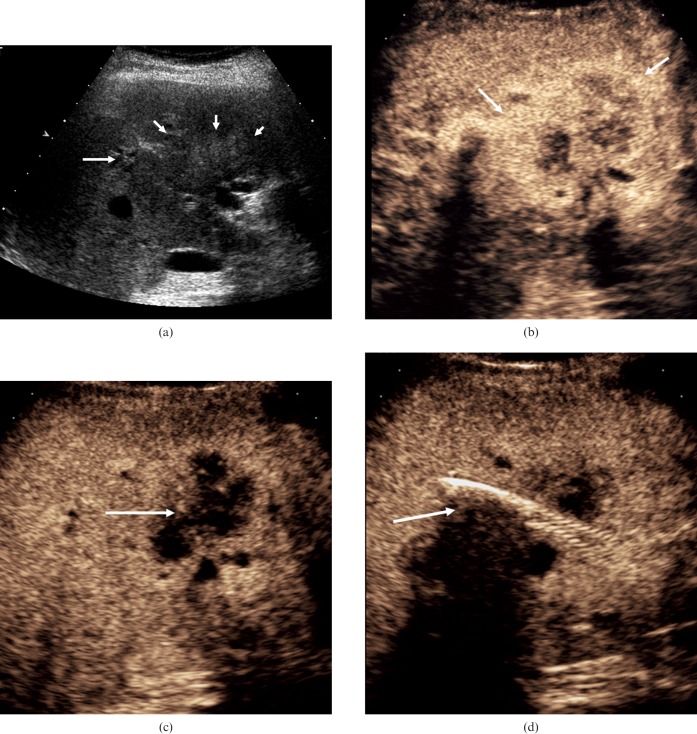
Figure 11.
Cholangiocarcinoma. (a) Baseline US illustrates an ill-defined echogenic heterogeneous mass at the porta hepatis (short arrows) with proximal dilatation of the intrahepatic bile ducts (long arrow). (b) CEUS at 20 s after microbubble contrast administration demonstrates peripheral irregular rim-like enhancement (arrows). (c) CEUS at 87 s after microbubble contrast administration demonstrates absence of enhancement within the tumour (arrow). (d) A metallic stent has been placed in the common bile duct for palliative reasons but is not functioning adequately. The CEUS image at 110 s demonstrates evidence of tumour invasion into the biliary stent (arrow).

