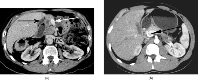
Figure 3.
(a) Axial post-contrast CT demonstrating preduodenal portal vein (white arrow), a dilated common bile duct proximal to the preduodenal portal vein (black arrow) and left-sided inferior vena cava (IVC). (b) Axial maximum intensity projections of a normal control volunteer showing the portal vein (black arrow) relationship to the first part of the duodenum (*). Note the portal vein traversing posterior to the first part of duodenum to enter the porta hepatis.

