Figure 4.

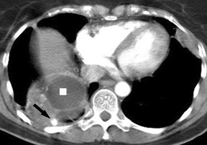
Follow-up CT scan from February 2008 demonstrating almost complete resolution of the left lung mass and significant regression in the right lower lobe mass, which still presents with varicosities (arrow) and cystic degeneration (square).

Follow-up CT scan from February 2008 demonstrating almost complete resolution of the left lung mass and significant regression in the right lower lobe mass, which still presents with varicosities (arrow) and cystic degeneration (square).