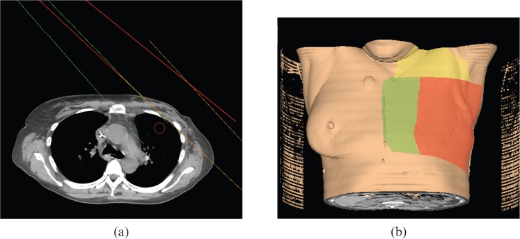
Figure 3.
Depiction of five-field arrangement. (a) Axial CT slice: red, medial tangent; orange, lateral tangent; green, angled IM (internal mammary) electron beam. (b) Three-dimensional depiction: red, surface path of medial and lateral tangents; yellow, surface path of anteroposterior (AP) and posteroanterior (PA) supraclavicular beam; green, surface path of angled IM electron beam.

