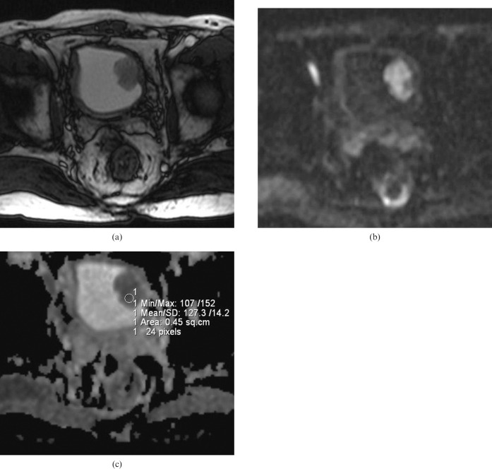
Figure 1.
(a) Axial T2 weighted MR image in a 64-year-old male depicting a 4 cm polypoid solid mass with a lobulated contour on the left lateral wall of the bladder. (b) On b = 1000 diffusion-weighted imaging, the mass shows a hyperintense signal corresponding to a restriction in diffusion. (c) Apparent diffusion coefficient (ADC) map of the same lesion. The ADC value of the lesion was measured as 1.27 × 10⁻³ mm2 s–1. Histopathologically, the lesion was reported as low-grade non-invasive urothelial carcinoma.

