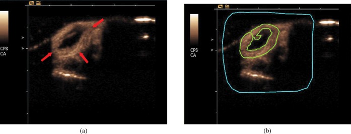
Figure 1.
(a) Transverse ultrasound images of an H22 tumour (arrow) obtained at peak enhancement after contrast agent injection. (b) Representative example of placement of a region of interest (ROI) (green line) drawn along the perimeter of each tumour and free from non-enhanced area. The region of blue line is a delimitative region used for motion compensation.

