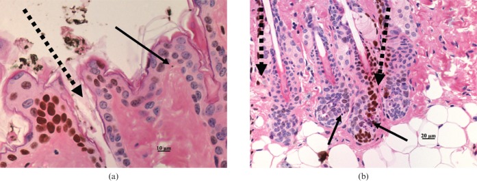
Figure 3.
Microbeam radiotherapy (MRT)-irradiated mouse skin stained with haematoxylin and eosin and the γ-H2AX immunohistochemical assay. (a) Apoptotic cell in “valley” region of epidermis; (b) apoptotic cell in both irradiated and non-irradiated hair follicles. Continuous arrows indicate apoptotic cells, dashed arrows indicate the path of the microbeams as inferred by γ-H2AX immunostaining.

