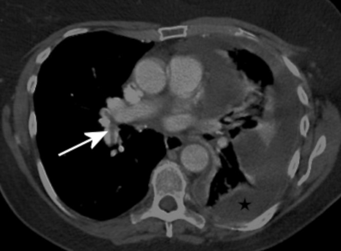
Figure 2.

Contrast-enhanced CT with 150 ml of contrast infused at 2.5 ml s–1. This shows a left-sided malignant pleural collection (star). There is sufficient enhancement of the pulmonary arteries to diagnose pulmonary emboli in the right lobar arteries (arrow).
