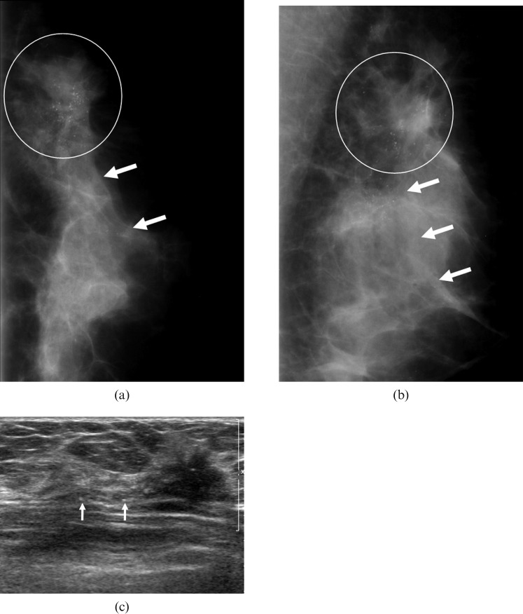
Figure 2.
A 45-year-old woman in whom screening at a local clinic had detected a mammographic abnormality in the left breast. (a,b) Mammograms show an irregularly shaped isodense mass with indistinct margin (circle), which is associated with segmentally distributed calcifications (arrows) in the upper outer quadrant of the left breast. (c) The sonogram shows an irregularly shaped hypoechoic mass with an indistinct margin in the left breast, which is associated with calcifications in or out of a mass. Surgery confirmed an invasive ductal carcinoma with high histological grade and positive lymphovascular invasion and extensive introductal component. This mass was also associated with negative oestrogen receptor status and positive HER-2/neu status.

