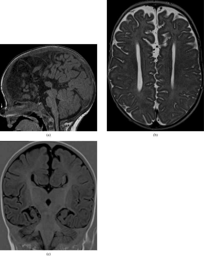
Figure 4.
6-year-old boy with corpus callosum (CC) agenesis: in the sagittal plane in MRI there is no structure consistent with the CC present in its usual location (a); a dermoid cyst of the nose associated with this malformation is observed (arrow). MR axial image: parallel lateral ventricles and “B bundles of Probst” (b). MR coronal image: “trident” anterior horns resembling “Viking helmet” or “mouse head” (c).

