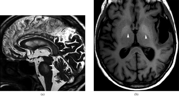
Figure 10.
47-year-old woman with known history of alcohol abuse presenting central pontine and extrapontine myelinolysis, attributed to rapid correction of hyponatraemia. MRI shows high signal in T2 images and low signal in T1 images at the level of the pons and the corpus callosum splenium (a and b, respectively), associated with spontaneously high signal in the globus pallidus (b, arrows) on T1 weighted images without contrast-media enhancement.

