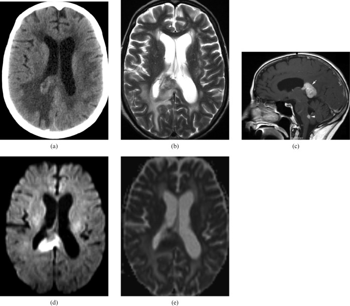
Figure 13.
66-year-old woman with lymphoma: non-contrast CT scan showing a spontaneously hyperdense mass at the corpus callosum (CC) splenium surrounded by extensive oedema (a). MRI is hyperintense in T2 images (b), with homogeneous enhancement at the CC splenium and the inferior part of the fourth ventricle (c, arrows). In diffusion weighted images (d, e) there is a restriction of diffusion enabling differential diagnosis from glioblastoma. A lesion with the same characteristics is also present in the brain stem as seen in the sagittal plane.

