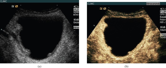
Figure 6.
62-year-old man with multiple bladder cancers. (a) Baseline ultasound (US) showed three polypoid lesions on the lateral bladder walls larger than 5 mm. (b) Contrast-enhanced (CE) ultrasound at 19 s showed the same number of polypoid lesions of different sizes extending into the bladder lumen. At cystoscopy two more lesions smaller than 5 mm, not identified using ultrasound or CEUS, were detected.

