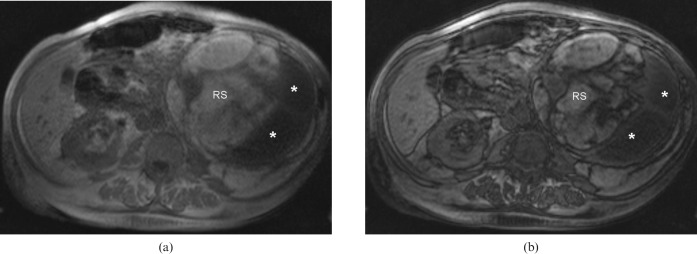
Figure 2.
Axial in-phase (a) and out-of-phase (b) gradient-echo T1 weighted images of the abdomen demonstrate T1 hyperintensity of the tissue expanding the left renal sinus (RS). Although the T1 hyperintensity by itself is non-specific, the peripheral etching along its margin with adjacent renal parenchyma indicates the fatty nature of this tissue. The dilated renal calyces peripherally are T1 hypointense (*), consistent with their fluid content.

