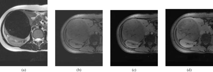
Figure 2.
MRI findings of a calcifying fibrous tumour of the liver. (a) T2 weighted image (T2WI) showing a dark signal-intensity mass in the right lobe of the liver. (b) Pre-contrast fat-suppressed T1 weighted image (T1WI) showing a lower signal-intensity mass corresponding to the T2WI. (c) Arterial and (d) 5 min-delayed contrast-enhanced T1WI showing a progressive delayed contrast-enhancement pattern of the mass similar to that of CT findings.

