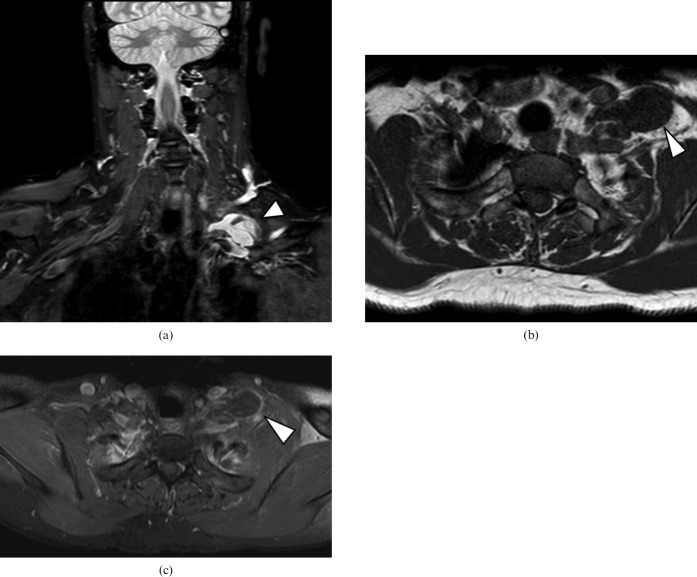
Figure 1.
(a) Coronal short tau inversion recovery sequence: a cystic lesion (arrowhead) is identified in the left supraclavicular fossa. (b) Axial unenhanced T1 weighted sequence shows a hypointense cyst (arrowhead) with a “pedicle” extending posterior to the internal jugular vein and anterior to the subclavian artery. (c) In the axial contrast-enhanced T1 weighted sequence with fat suppression the lesion has an absence of enhancement and a location consistent with a lymphocoele (arrowhead) of the thoracic duct termination.

