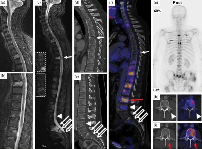
Figure 1.
(a,b) Outpatient MRI of the spine (T2 weighted turbo inversion recovery magnitude (TIRM) FS) showing vertebrae C1–T5 (a) and T6–L2 (b) prior to radiation therapy. Multiple hyperintense lesions representing vertebral metastases to T3, T7, T11–12, L1, L3, S1–2 and a compression fracture of T7 protruding into the spinal canal are visible. (c) MRI of the spine after radiotherapy (T2 weighted TIRM fat-saturation (FS); insets, T1 weighted turbo spin-echo (TSE) with gadolinium intravenous contrast (top) and T1 weighted TSE (bottom) of L3–S1). Hyperintense lesions appear unchanged in radiated regions. (d,e) Sagittal CT of the thoracic (d) and lumbar (e) spine showing the compression fracture of T7 and inhomogeneous bone structure but no apparent osteolysis. (f) Positron emission tomography (PET)/CT fusion image of the vertebral column with previous biopsy sites (solid arrows, transpedicular biopsy of vertebrae T7 and S1; open arrows, surgical (open) biopsy of vertebral bodies S1 and S2; arrow heads, transpedicular biopsy of vertebra L4) and biopsy site chosen according to PET/CT (red arrow). (g) Bone scintigraphy showing increased uptake of 99mTc-DPD in several ribs on the left and right hemithorax, as well as in the vertebral column. (h) PET/CT fusion image of L4: the biopsy channel is visible adjacent to FDG-avid, potentially vital malignant tissue. (i) Retrospective fusion of PET data (right) with post-biopsy CT (left) after PET/CT-planned biopsy of vertebra L3.

