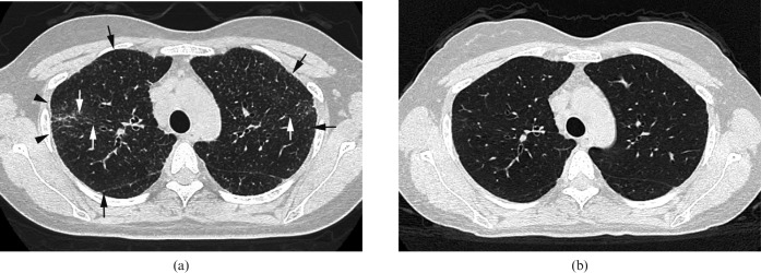
Figure 3.
Images in a 35-year-old woman. (a) Transverse CT scan (1 mm section thickness) obtained at the level of upper lobes shows multiple small nodules (arrows) and architectural distortion (arrowheads; KL-6, 1000 U ml–1). Traction bronchiectasis and bronchial wall thickening are also present (white arrows). (b) Transverse CT scan (1 mm section thickness) obtained 6 months later demonstrates disappearance of nodules, architectural distortion, traction bronchiectasis and bronchial wall thickening (KL-6, 285 U ml–1).

