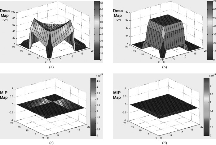
Figure 8.
Graphs showing dose and malignant induction probability (MIP) maps for a two-field right-angled wedged pair arrangement for X-rays (a) and two-field protons (b) when the planning target volume is moved towards the corner of the overall volume under consideration. Note the three orders of magnitude difference in the MIP scale which vary from multiplication factors of 10−17 for X-rays and 10−20 for protons. The total MIPs are protons 0.11, X-rays 0.25.

