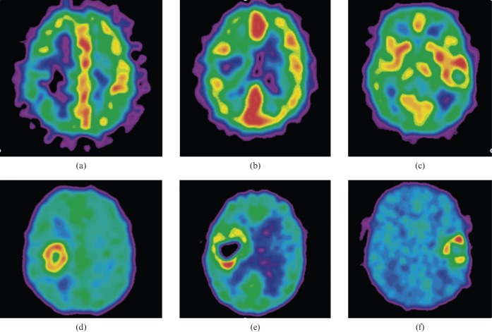
Figure 1.
(a–c) 15O-H2O positron emission tomography (PET) perfusion images in three patients with glioblastoma multiforme. (d–f) Corresponding late 18F-labelled fluoro-misonidazole (18F-MISO) PET images show tumour hypoxia in low perfusion, in intermediate perfusion with an inverse pattern compared with hypoxia and in high perfusion. PET images are normalised to their own maximum. Reproduced with permission from [119].

