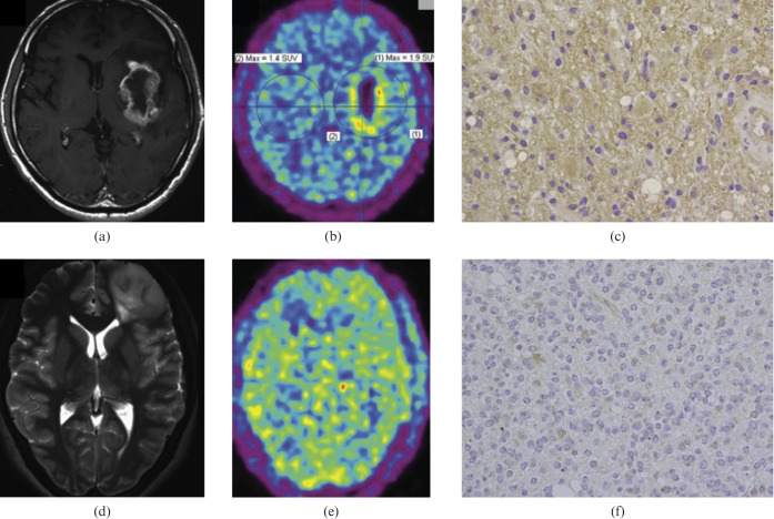
Figure 3.
(a–c) A 68-year-old man with a glioblastoma multiforme (GBM). Axial T1 weighted gadolinium-enhanced MR image (a) showing ring enhancement in the left insula, FRP-170 positron emission tomography (PET) image (b) showing marked uptake, and photomicrograph with hypoxia inducible factor-1α (HIF-1α) antibody (c) indicating strong immunoreactivity. Axial T2 weighted MR image (d) showing a diffuse infiltrative lesion in the left frontal lobe, FRP-170 PET image (e) showing moderate uptake within the lesion, and photomicrograph with HIF-1α antibody (f) showing moderate immunoreactivity. Reproduced with permission from [139].

