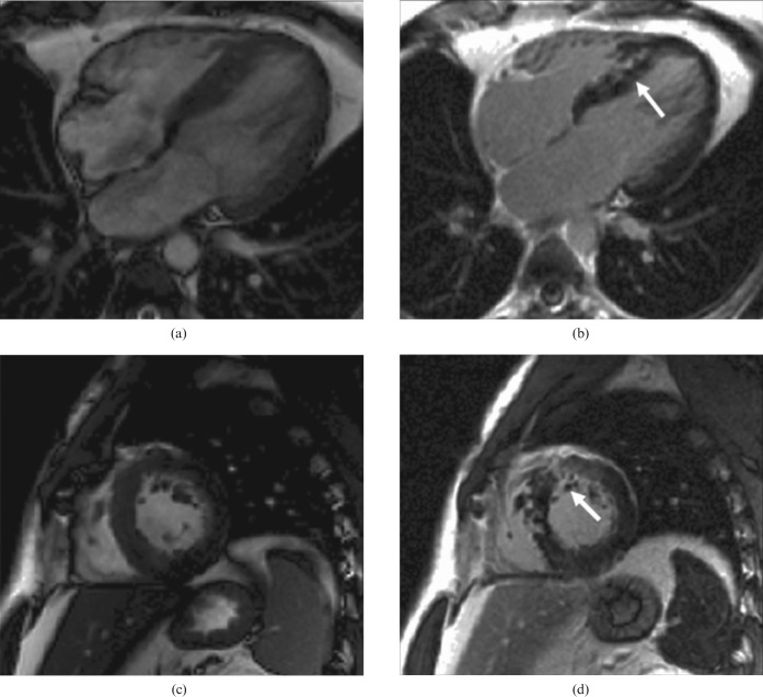
Figure 1.
Hypertrophic cardiomyopathy. Note left ventricular hypertrophy and, after contrast, late gadolinium enhancement of the hypertrophied areas and right ventricular insertion points (arrows). (a, c) Steady-state free precession cine in diastole [(a) four-chamber view; (c) short-axis view]. (b, d) Inversion recovery after gadolinium bolus [(b): four-chamber view; (d) short-axis view].

