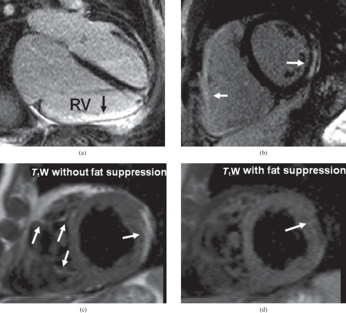
Figure 8.
Arrhythmogenic right ventricular (RV) cardiomyopathy: note the dilated right ventricle (larger than the left ventricle) which had impaired systolic function on cine imaging. (a) Inversion recovery after gadolinium, four-chamber view, shows enhancement of the RV free wall. (b) Inversion recovery after gadolinium image confirms the RV free wall late gadolinium enhancement and shows left ventricular involvement with mid-ventricular inferolateral subepicardial enhancement. (c, d) T1 weighted (T1W) images without (c) and with (d) fat suppression from another patient with arrhythmogenic right ventricular cardiomyopathy shows fat infiltration in the right and left ventricles (arrows).

