
Figure 7.
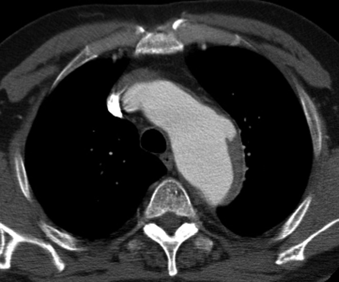
This patient, who has several comorbidities, presented with chest pain. Only a post-contrast study was performed, which shows a focal out-pouch of contrast into the atheroma in the distal arch typical of a penetrating atherosclerotic ulcer. Some of the aortic wall thickening resolved on subsequent follow-up CT angiogram making it likely to have been an intramural haematoma (IMH). A non-contrast-enhanced CT would have helped by showing the acute IMH as a hyperdense rim.