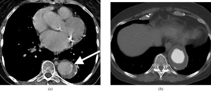
Figure 9.
Two CT angiograms on different patients demonstrating the difficulty differentiating extensive intramural haematoma (IMH) from intraluminal thrombus in the aorta. (a) A patient with an extensive IMH involving the descending aorta. The clue that this is not simply intraluminal thrombus is the displacement of intimal calcification (arrow). (b) A different patient in whom there is intraluminal thrombus accumulating in an ectatic section of the descending aorta. The intimal calcification is not displaced. Sometimes chronic intraluminal thrombus can develop calcification within it, which can make it more difficult to differentiate.

