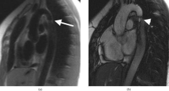
Figure 12.
Coarctation in a 19-year-old female with hypertension. (a) MRI “black blood” half-Fourier acquisition single-shot turbo spin echo sequence shows the anatomical narrowing (arrow). (b) Image from a cine balanced steady-state free precession sequence demonstrates the stenotic jet (arrowhead). Performing a phase contrast study perpendicular to this enables the peak velocity and subsequently the gradient across the stenosis to be estimated.

