
Figure 13.
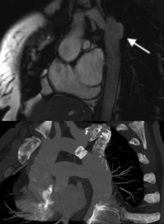
Coarctation repair complications. (Top figure) Sagittal oblique balanced steady-state free precession image demonstrating aneurysm formation in a 29-year-old woman, which is a common complication of synthetic patch repair (arrow). (Bottom figure) Sagittal oblique maximum intensity projection in a 41-year-old man with a recurrent coarctation which was initially treated with a bypass graft. The graft eventually became calcified and stenosed (after many years), so a stent was placed in the original native coarctation to relieve the obstruction.