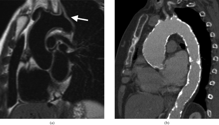
Figure 14.
Two patients with Takayasu's disease. (a) Sagittal oblique half-Fourier acquisition single-shot turbo spin echo “black blood” image which is the ideal sequence for demonstrating the diffuse thickening of the aortic wall and branches for the arch (arrow). (b) Contrast-enhanced CT angiogram sagittal oblique multiplanar reformat image of a patient with chronic disease with a diffusely calcified aorta.

