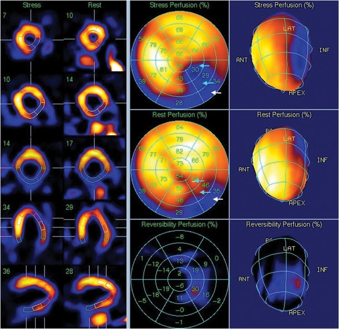
Figure 1.
Rest and stress images from a cardiac nuclear medicine myocardial imaging study. The images demonstrate a severe perfusion defect in the inferolateral wall. This is partly fixed (in the basal segment; white arrows) and partly reversible (in the distal and mid-inferolateral wall; cyan arrows) and is typical for an area of ischaemia.

