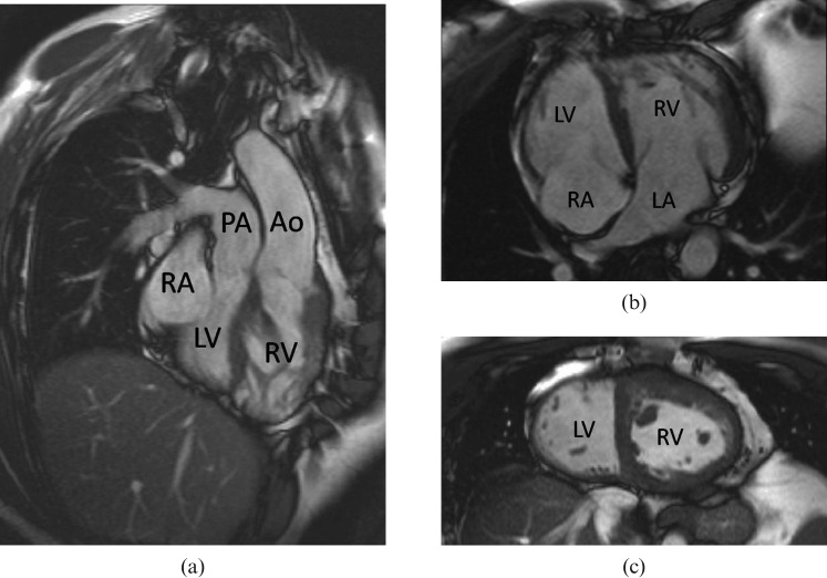
Figure 4.
Unoperated “congenitally corrected” transposition of the great arteries shown by MRI. (a,b) Both the atrio-ventricular and the ventriculo-arterial connections are discordant. Note the expected apical displacement of the septal insertion of the tricuspid valve of the right ventricle (RV) relative to that of the mitral valve of the left ventricle (LV), and the hypertrophied muscle of the systemic RV. (c) In the mid short axis image, the left ventricular cavity can be identified as the one on the smoother, less trabeculated, side of the ventricular septum. PA, pulmonary artery; Ao, aorta.

