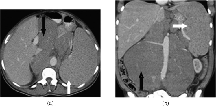
Figure 2.
Images from a 65-year-old male with relapse of small lymphocytic lymphoma. (a) Axial and (b) coronal contrast-enhanced CT images reveal moderate hepatosplenomegaly with multiple low attenuation, subcentimetre micronodules in the spleen (white arrows). Associated bulky retroperitoneal (black arrows) and mesenteric lymphadenopathy are also seen.

