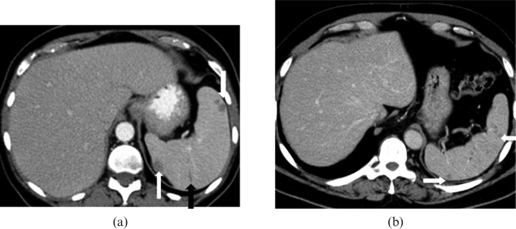
Figure 11.
Images from a 65-year-old male with acute myelogenous leukaemia. (a) Axial contrast-enhanced CT image reveals mild hepatosplenomegaly with multiple well-defined low attenuation, non-enhancing, <1 cm micronodules (white arrows) in the spleen due to leukaemic deposits. An associated small linear area of splenic infarct (black arrow) is also noted. (b) Axial contrast-enhanced CT image of a 60-year-old patient with clinical suspicion of fungal microabscesses reveals multiple well-defined low attenuation, minimally enhancing, subcentimetre nodules (white arrows) in the normal-sized spleen making it difficult to differentiate from leukaemic lesions; however, hyperintense signal on T2 weighted images with rim enhancement on MRI (not shown) were suggestive of fungal microabscesses.

