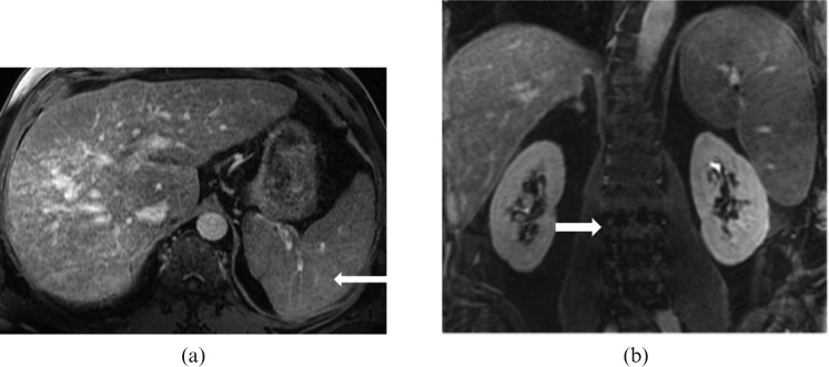
Figure 15.
Images from a 59-year-old male with relapsed refractory IgG myeloma. (a) Axial T1 post-contrast venous phase and (b) coronal T1 post-contrast delayed phase MRI reveal moderate hepatosplenomegaly that was histopathologically proved to be a result of a combination of cirrhosis and multiple myeloma. Note the multiple tiny hypointense nodules in the spleen representing Gamna–Gandy bodies (long white arrow) due to underlying cirrhosis of liver with portal hypertension. Note the heterogeneous appearance of bone marrow of the lumbar spine (short white arrow) secondary to multiple myeloma.

