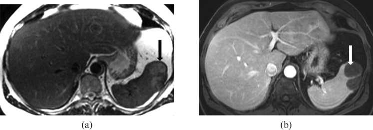
Figure 17.
Images of a 63-year-old male with chronic myeloid leukaemia and extramedullary haematopoiesis. (a) Axial T2 and (b) T1 axial dynamic venous MRI reveals a normal-sized spleen with a focal lobulated lesion in its anterior aspect appearing heterogeneously hyperintense on T2 weighted images (black arrow), hypointense on T1 (not shown) and with minimal enhancement on post-gadolinium fat suppressed gradient recalled echo T1 weighted image (white arrow).

