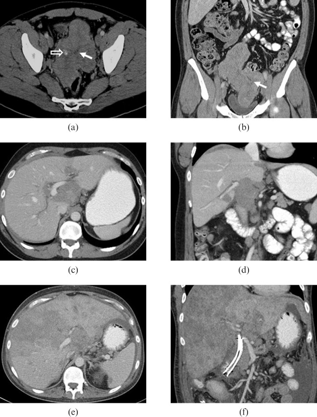
Figure 2.
29-year-old male presented with right upper quadrant discomfort. Initial contrast-enhanced (a) axial and (b) coronal CT images show a large heterogeneous lobulated mass in the pelvis with foci of calcification (open arrow) and multiple soft-tissue masses within the pelvis and mesentery. Central stellate low density area corresponds to an area of fibrosis (solid arrows). As the disease progressed the patient developed biliary obstruction and intrahepatic biliary ductal dilation as a result of metastatic disease in the porta hepatis (c,d) and then extensive hepatic metastatic disease (e,f). The patient died 27 months after initial diagnosis.

