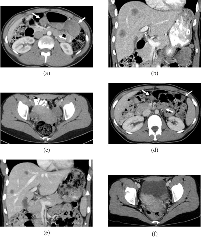
Figure 3.
An otherwise healthy 26-year-old female presented with enlarged right external iliac lymph nodes. (a–c) Initial contrast-enhanced CT shows a dominant mass in the rectouterine space inseparable from the uterus and multiple smaller intraperitoneal soft-tissue masses, enlarged retroperitoneal lymph nodes and metastatic disease in the liver. (d–f) After four cycles of chemotherapy the lesions are significantly decreased in size. Arrows indicate representative lesions.

