Figure 3.
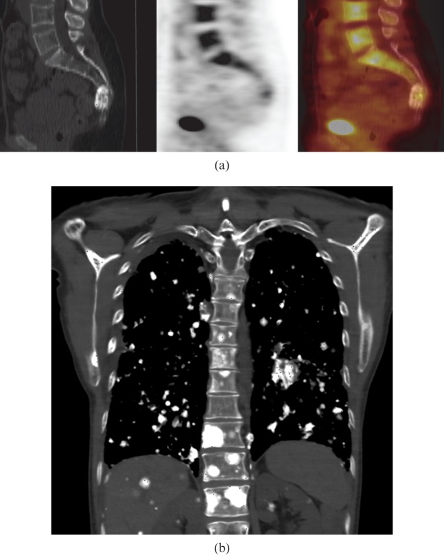
54-year-old female with primary sacral osteosarcoma. (a) Sagittal 18F-fluorodeoxyglucose (18F-FDG) positron emission tomography/CT images showing mild 18F-FDG uptake in a sclerotic mass involving the fourth sacral vertebra at presentation. This is not typical of osteosarcomas, which usually demonstrate more FDG uptake. (b) Coronal CT of the chest shows multiple calcified nodules in lungs, liver, kidneys, ribs, right scapula and thoracic vertebra, in keeping with diffuse metastatic osteosarcoma 3 years after presentation.

