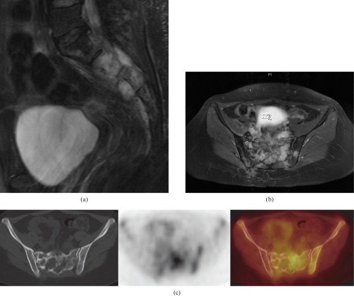
Figure 4.
25-year-old female with recurrent primary clear cell sarcoma of the sacrum. (a) Sagittal short-inversion-time inversion recovery MR image showing a hyperintense mass involving the second and third sacral vertebra with extension of the mass anteriorly into the pre-sacral space and also posteriorly. (b) Axial T1 weighted fat-suppressed contrast-enhanced MR images showing enhancement of the mass with intracanalicular extension. (c) Axial 18F-fluorodeoxyglucose (18F-FDG) positron emission tomography/CT images showing a mixed lytic and sclerotic lesion of the sacrum. Increased 18F-FDG uptake is asymmetrical, with more uptake on the left correlating with the site of less severe bone destruction.

