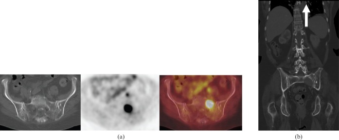
Figure 8.
75-year-old female with sacral lytic bone metastasis from primary non-small cell lung cancer. (a) Axial 18F-fluorodeoxyglucose (18F-FDG) positron emission tomography/CT images showing a lytic lesion in the left sacrum, which is 18F-FDG-avid. (b) Coronal CT of the abdomen and pelvis 6 months later showing a larger lytic lesion in the left sacrum with a soft tissue component. Note the surgical suture material in the left infrahilar region from previous left lower lobectomy (arrow).

