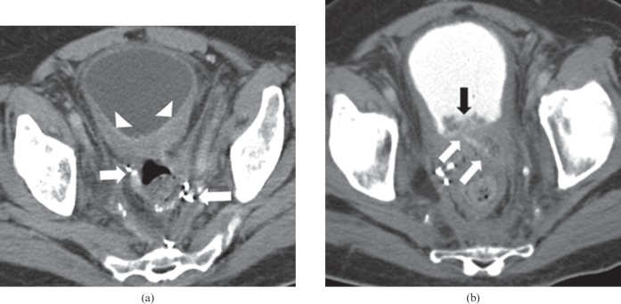
Figure 9.
68-year-old male with rectal carcinoma, post-low anterior resection and adjuvant radiation. (a) Pre-treatment axial contrast-enhanced CT of the pelvis demonstrates thickening of the posterior wall of the urinary bladder (arrowheads), which is probably radiation associated. Arrows point to surgical anastomosis from the previous anterior resection. The patient was treated with bevacizumab, fluorouracil and leucovorin when he developed haematuria, pneumaturia and fecaluria during treatment. (b) Post-treatment axial contrast-enhanced CT cystogram, with retrograde contrast instillation into the urinary bladder via a Foley catheter (not seen), shows contrast flowing along the fistulous tract from the urinary bladder to the rectum (white arrows). New irregular soft tissue thickening of the posterior wall of the urinary bladder is present (black arrow). Chemotherapy and bevacizumab were discontinued. The patient was treated with antibiotics. However, following recurrent episodes of urosepsis he required a diverting end colostomy.

