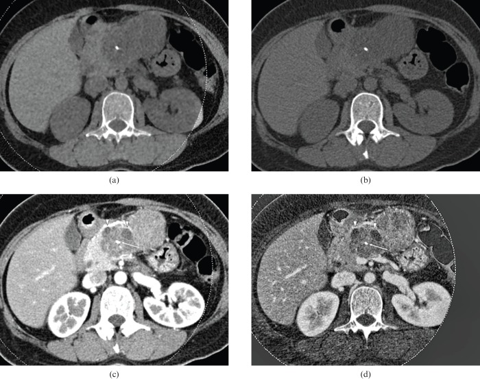
Figure 2.
40-year-old female with mucinous cystic adenoma. Dual-energy CT images with a weight factor of (a) 0.3, (b) 0.5, and (c) 0.7, all show a large, cystic mass with the thin wall as well as internal septation in the pancreas tail. The image with a weight factor of 0.3 shows the least noise, although the septation is better visualised as internal septation is on the weight factor 0.7 image. Note that the internal cystic component appears to be coarse on the image with weight factor 0.7. The image with weight factor 0.5 shows relatively higher contrast than the image with weight factor 0.3 and lower noise than the images with weight factor 0.7. Both reviewers selected image (b) as the preferred image.

