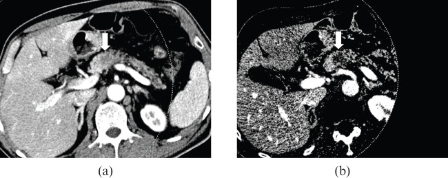
Figure 3.
55-year-old male with pancreas body adenocarcinoma. (a) The axial CT scan with a weight factor 0.3 shows an ill–defined, low-attenuation lesion (arrow) in the pancreas body. (b) The iodine map shows much better lesion conspicuity compared with the mixed image, due to the higher lesion-to-pancreas contrast.

