Figure 1.
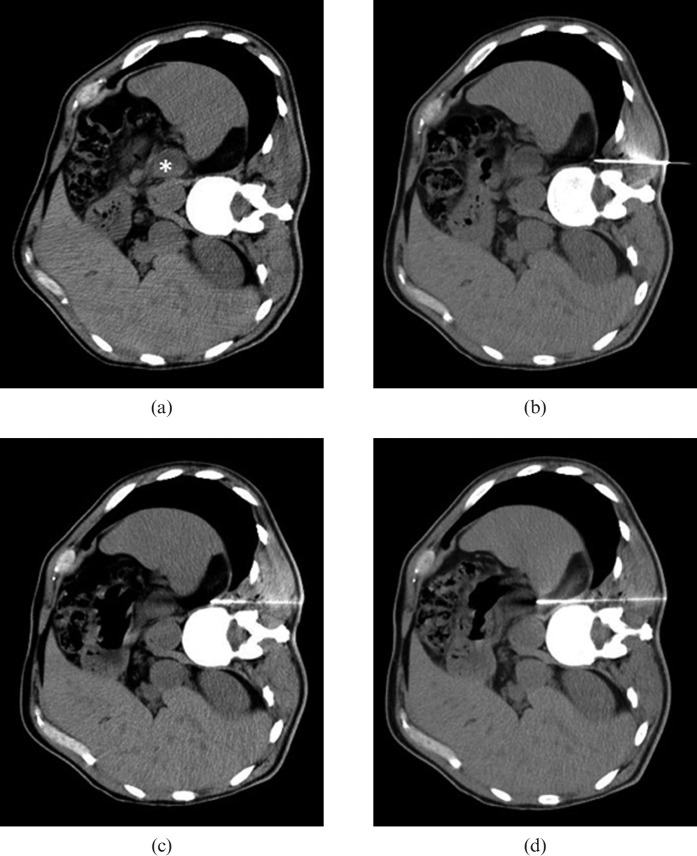
Example of CT-guided paravertebral biopsy of the left adrenal gland using hydrodissection. (a) Axial CT of the abdomen with the patient in right lateral decubitus without the use of intravenous contrast, showing hypodense nodule in the left adrenal gland (*). (b) The tip of a 17-gauge coaxial needle was positioned in the fat plane between the vertebral body and the left parietal pleura. (c) After the injection of 20 ml of 0.9% saline solution, parietal pleura was displaced, expanding the posterior paravertebral space while simultaneously advancing the coaxial needle carefully under serial CT control. (d) During suspension of respiration, the coaxial needle tip traversed the diaphragm and was positioned next to the left adrenal gland. Five fragments were then obtained using an 18-gauge needle.

