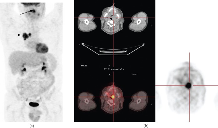
Figure 3.
53-year-old male scanned to assess the resectability of a known squamous cell carcinoma. (a) Coronal positron emission tomography (PET) scan shows a right hilar lung mass (lower arrow) and unexpected oropharyngeal 18-fludeoxyglucose (FDG) tracer uptake (upper arrow). (b) Transaxial PET/CT shows intense FDG uptake within a soft-tissue mass within the left side of the oropharynx. Follow-up examination revealed tonsillitis.

